A Small Beautiful World
Scale is an interesting thing because we don’t know where we sit on the, well, scale. It’s easy to take a self-centered solipsistic approach and measure everything in terms of our own perspective. It has its practical applications after all. Things are bigger or smaller than you. Some things are dangerously big, on the other hand some things are dangerously small and probably airborne in a public space. You’d think that the smaller things get, the less complex they are. Instead we look through our microscopes and see entire worlds crawling with life. Beauty in a micron, a Fabergé egg of mysteries. Then we lean back and look up to the flakey plastered ceiling above us and the sky beyond it. We might wonder then, is someone looking down at us right now? Is our entire human history contained within a falling raindrop? Perhaps our Milky Way is a collection of atoms holding together a tiny bit of a chair leg for giants. While these are questions we’ll never be able to answer, we can at least appreciate the beauty of the microscopic world.
Many of us don’t have easy access to the necessary tools, but the winners of the 48th Annual Nikon Small World Photomicrography Competition have us covered. Their photos have revealed and celebrated the beauty that lies all around us in one form or another. The skills required to capture these moments are multidisciplinary, with a firm grasp of technological processes as well as the artistic flair to create an aesthetic shot.
This year’s first place prize was awarded to Grigorii Timin, who used high-resolution microscopy to capture an embryonic hand of a Madagascar giant day gecko.
“This embryonic hand is about 3 mm (0.12 in) in length, which is a huge sample for high-resolution microscopy,” said Timin. “The scan consists of 300 tiles, each containing about 250 optical sections, resulting in more than two days of acquisition and approximately 200 GB of data.”
He went on to say, “The Nikon Small World Competition is a great opportunity to share how impressive nature is on a microscopic level, not only within a scientific community but also with the general public.”
“Each year, Nikon Small World receives an array of microscopic images that exhibit exemplary scientific technique and artistry. This year was no exception,” said Eric Flem, Communications and CRM Manager, Nikon Instruments. “At the intersection of art and science, this year’s competition highlights stunning imagery from scientists, artists, and photomicrographers of all experience levels and backgrounds from across the globe.”
The following images are among the top 12 from this year’s competition.
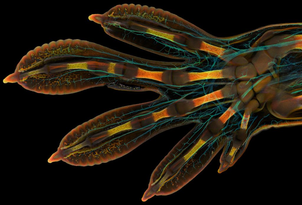
1st. Photographer: Grigorii Timin & Dr. Michel Milinkovitch | Location: Geneva, Switzerland
Image Description: Embryonic hand of a Madagascar giant day gecko (Phelsuma grandis). Confocal, 63X (Objective Lens Magnification).
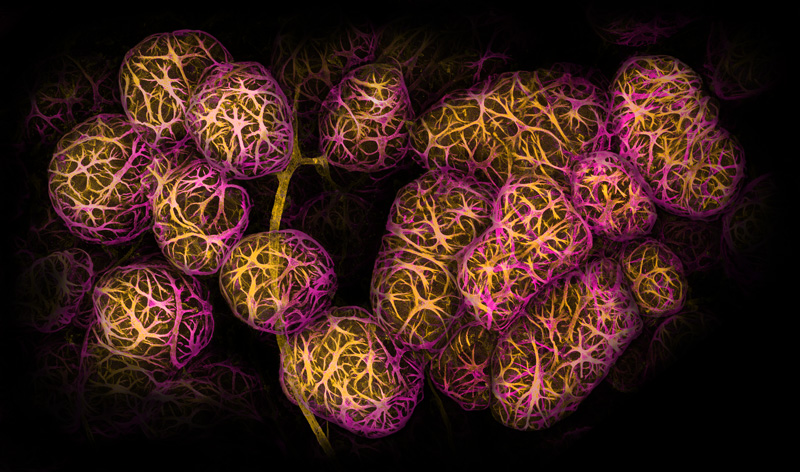
2nd. Photographer: Dr. Caleb Dawson | Location: Melbourne, Victoria, Australia
Image Description: Breast tissue showing contractile myoepithelial cells wrapped around milk-producing alveoli. Confocal, 40X (Objective Lens Magnification).
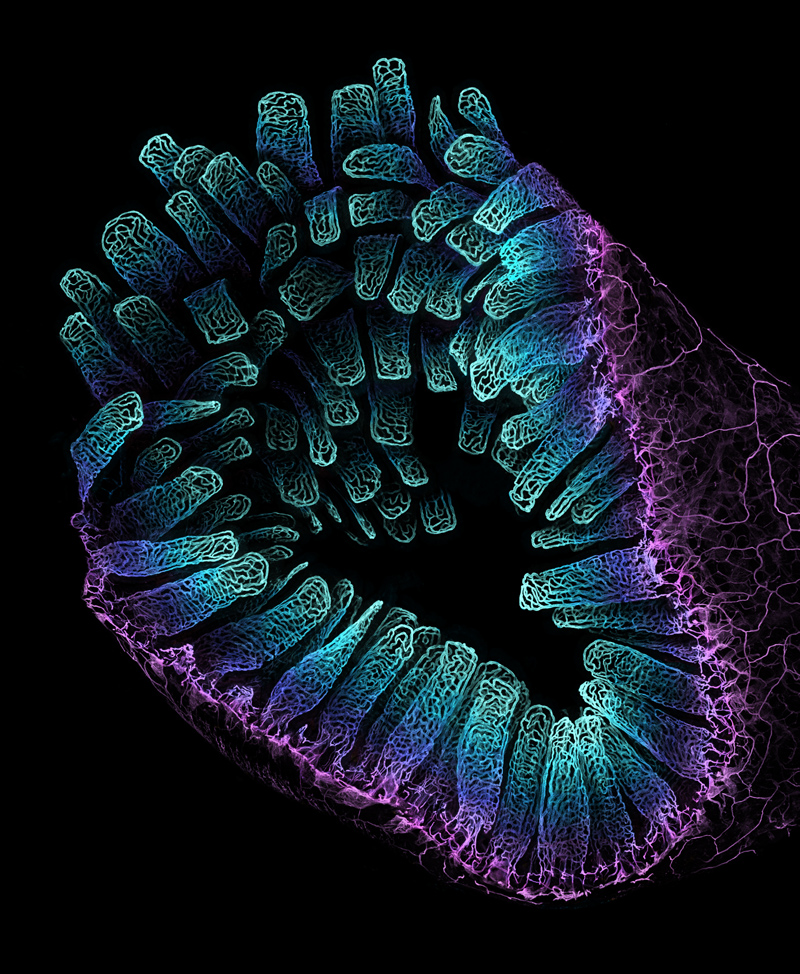
3rd. Photographer: Satu Paavonsalo & Dr. Sinem Karaman | Location: Helsinki, Finland
Image Description: Blood vessel networks in the intestine of an adult mouse. Confocal, 10X (Objective Lens Magnification).
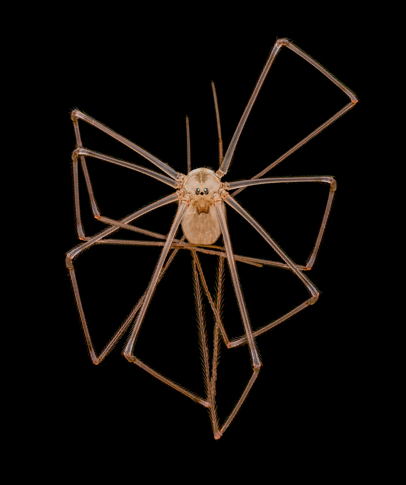
4th. Photographer: Dr. Andrew Posselt
Location: Mill Valley, California, USA
Image Description: Long-bodied cellar/daddy long-legs spider (Pholcus phalangioides). Image Stacking, 3X (Objective Lens Magnification).
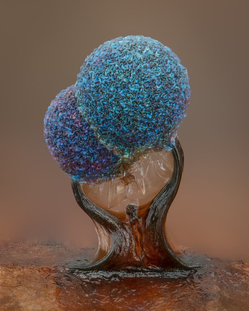
5th. Photographer: Alison Pollack
Location: San Anselmo, California, USA
Image Description: Slime mold (Lamproderma).
Image Stacking, Reflected Light,
10X (Objective Lens Magnification).
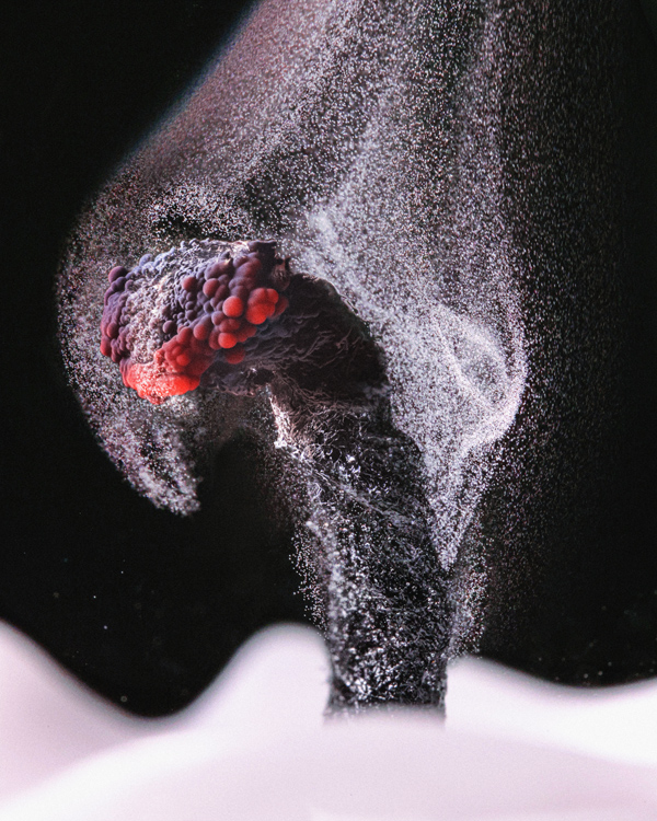
6th. Photographer: Ole Bielfeldt
Location: Cologne, North Rhine-Westphalia, Germany
Image Description: Unburned particles of carbon released when the hydrocarbon chain of candle wax breaks down Brightfield, Image Stacking, 2.5X (Objective Lens Magnification).
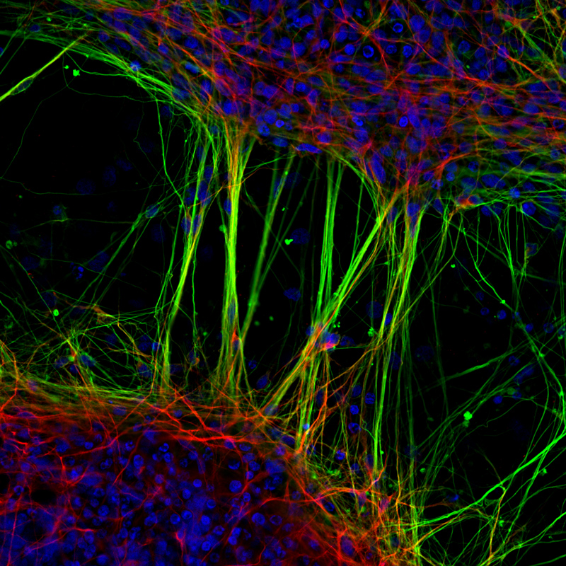
7th. Photographer: Dr. Jianqun Gao & Prof. Glenda Halliday Location: Sydney, New South Wales, Australia
Image Description: Human neurons derived from neural stem cells (NSCs). Confocal, Fluorescence, 20X (Objective Lens Magnification).
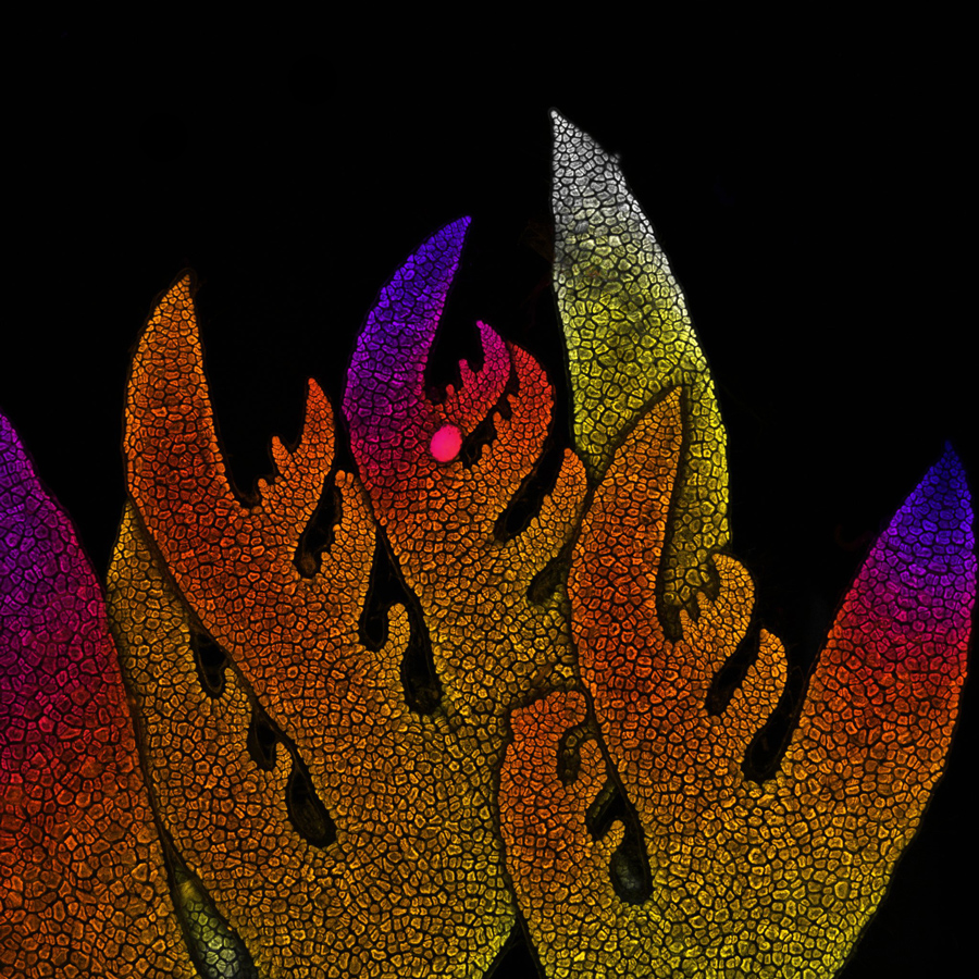
8th. Photographer: Dr. Nathanaël Prunet | Location: Chapel Hill, North Carolina, USA
Image Description: Growing tip of a red algae. Confocal, 10X (Objective Lens Magnification).
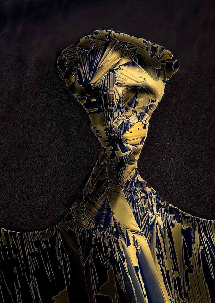
9th. Photographer: Dr. Marek Sutkowski | Location: Warsaw, Poland
Image Description: Liquid crystal mixture (smectic Felix 015). Image Stacking, Polarized Light, 40X (Objective Lens Magnification).
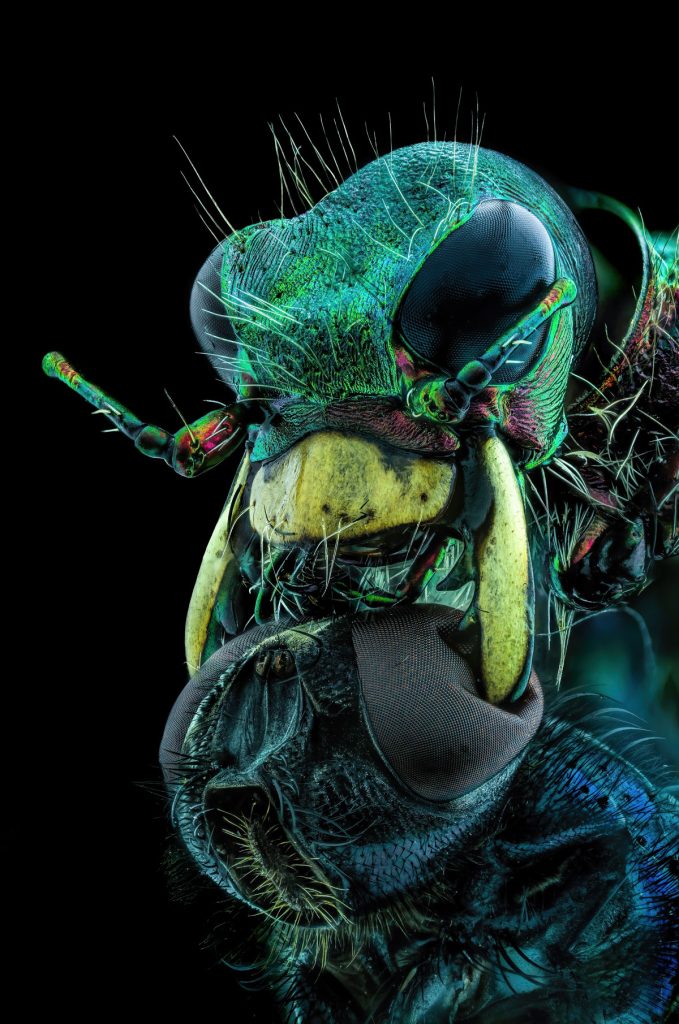
10th. Photographer: Murat Öztürk | Location: Ankara, Turkey
Image Description: A fly under the chin of a tiger beetle. Image Stacking, 3.7X (Objective Lens Magnification).
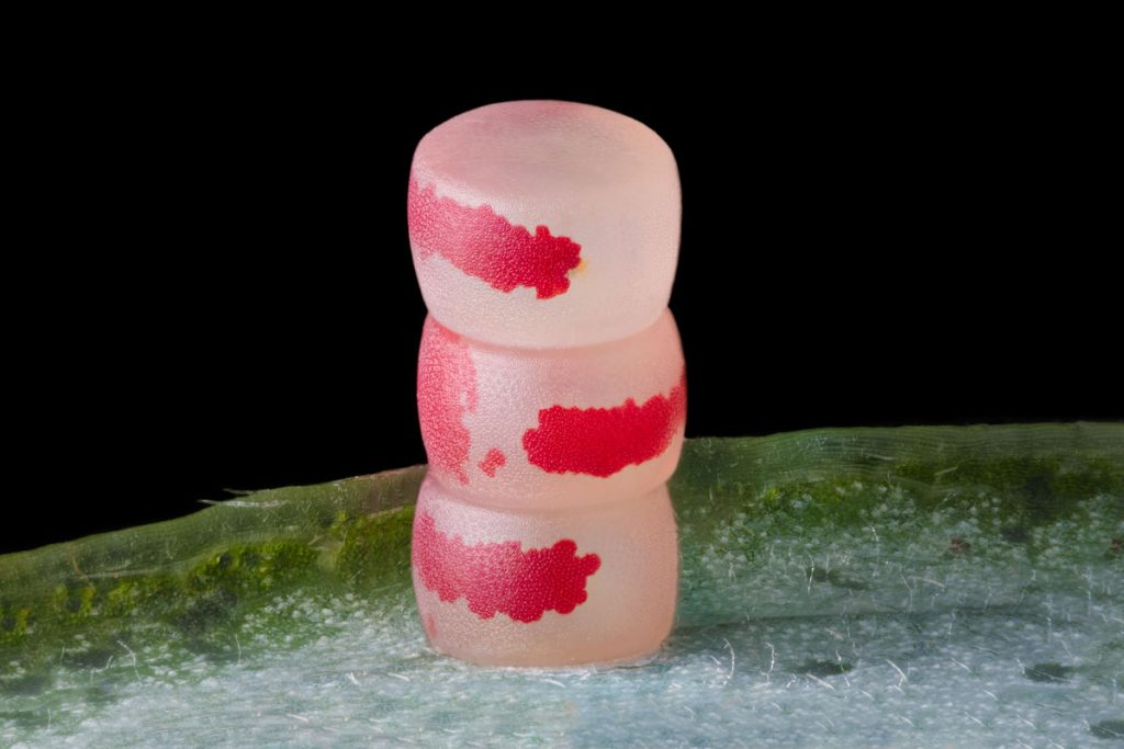
11th. Photographer: Ye Fei Zhang | Location: Jiang Yin, Jiangsu, China
Image Description: Moth eggs. Image Stacking, 10X (Objective Lens Magnification).
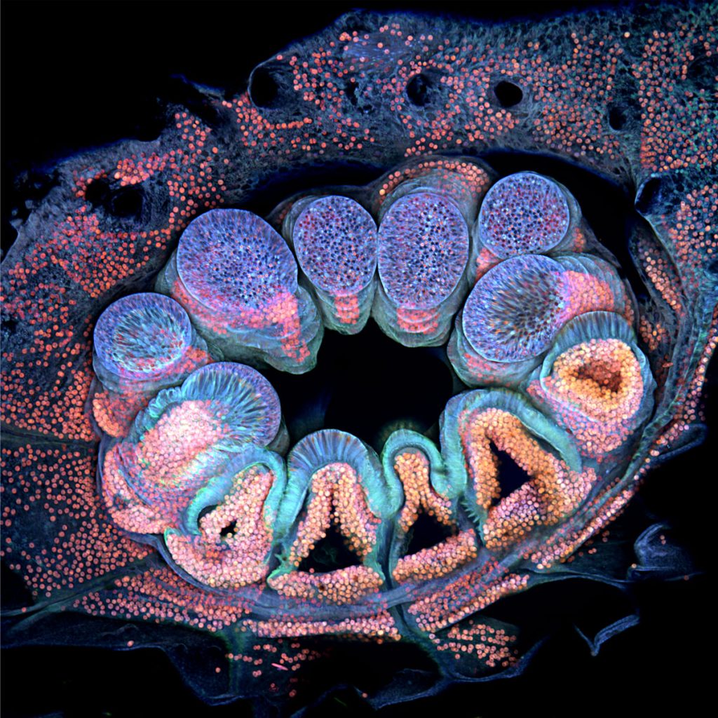
12th. Photographer: Brett M. Lewis
Location: Brisbane, Queensland, Australia
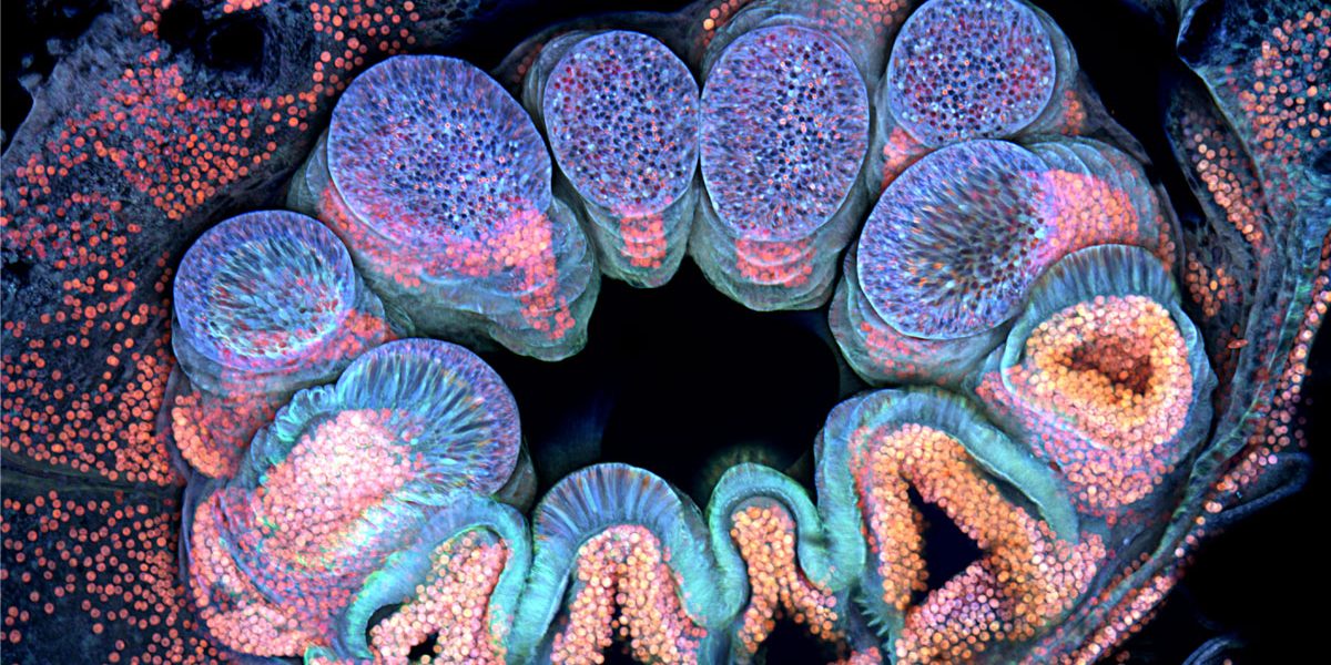
Image Description: Autofluorescence of a single coral polyp (approx. 1 mm). Fluorescence, Image Stacking, 20X (Objective Lens Magnification).
